
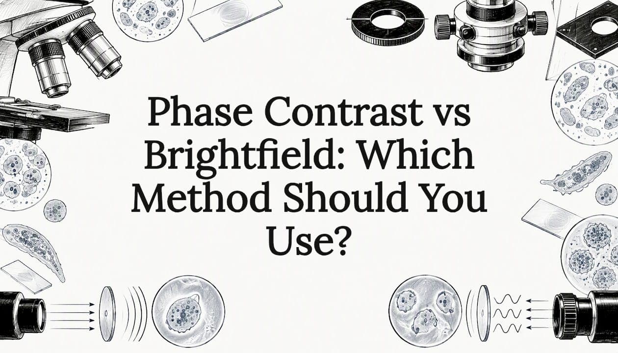
Brightfield microscopy is the standard method: light passes through the specimen and you see a dark image on a bright background. Phase contrast is a way of adding contrast to specimens that are transparent, such as living cells, without staining them. Choose brightfield for stained slides, general teaching and most home use. Choose phase contrast when you need to watch living, unstained cells in detail.
This guide explains how each method works, what each one is good at, where each one fails, and what equipment you need. You will also see how darkfield fits in and how to decide for your own bench or classroom.
TL;DR
- Brightfield shows dark, absorbing structures against a bright field. It works best on stained or naturally colored specimens, and it is simple, inexpensive and the default in classrooms.
- Phase contrast turns tiny differences in how light is delayed by a specimen into differences in brightness. It makes unstained living cells visible without killing them.
- Phase contrast needs extra parts: a condenser annulus and phase-contrast objectives with a phase plate. It also costs some resolution and brightness.
- Learn brightfield first, with correct Köhler illumination. Add phase contrast only if your work is live, unstained cells.
Table of Contents
- What brightfield microscopy does
- What phase contrast microscopy does
- Phase contrast vs brightfield at a glance
- Which specimens suit each method
- Where darkfield fits
- Get brightfield right before you add anything
- Our perspective
- Choosing a microscope for your needs
- FAQ
- Sources
What brightfield microscopy does
In brightfield, light from below is transmitted through the sample. According to OpenStax, the image appears dark against a bright background, and contrast comes from differences in how the specimen transmits, absorbs, reflects or refracts light.
That description tells you what brightfield is good at. Anything that absorbs light, such as a stained tissue section, pigmented algae, or a red blood cell, stands out clearly. Anything that absorbs almost nothing, such as a clear living cell in water, barely shows.
Advantages of brightfield
- Simple optics. You need a transmitted-light source, a condenser, an objective and an eyepiece or camera. The University of British Columbia's imaging facility notes that Köhler illumination is important for getting the best image.
- Full aperture. Nothing blocks part of the light path, so you get the resolution the objective was designed for.
- Bright, natural images. Color stains look like color stains, which matters for histology and microbiology teaching.
- Lower cost and less setup. Every compound microscope does it.
In a comparison of brightfield and phase-contrast imaging published in 2010, brightfield was found to be simpler and less expensive, and gave the best overall results in that study's application. Treat that as one study's finding, not a universal rule: the answer depends on your specimen.
Limitations of brightfield
The main limit is contrast. OpenStax notes that stains are commonly used to add contrast to colorless, transparent specimens, but staining can kill or alter living material. Transparent unstained specimens can be nearly invisible.
There is also a physical ceiling on useful magnification. Brightfield images stay sharp up to about 1000×; going beyond that adds size, not detail. Our guide on why 1000× is the limit for home microscopes and our explanation of resolution versus magnification cover this in detail.
What phase contrast microscopy does
Living cells are mostly water, so they absorb very little light. But they are not optically empty. Cell parts such as the nucleus and cytoplasm have a different refractive index than the water around them, so light passing through them is slightly delayed. Your eye cannot see that delay, which is a shift in phase.
Phase contrast converts that invisible shift into a visible difference in brightness. The technique was developed by Frits Zernike, and it is described as giving high-contrast images of unstained cells compared with brightfield.
How the optics work
Two parts do the job:
- A ring-shaped stop (annulus) in the condenser. It creates a hollow cone of illumination.
- A phase plate or phase ring in the objective. It makes the direct light and the light refracted by the specimen interfere, so small phase differences become differences in brightness.
The result is usually a gray image in which cell edges, nuclei and organelles appear darker or brighter than the background, depending on the type of phase plate. Our reading of the optics is that the annulus and the phase ring must line up exactly. That is why phase contrast microscopes include a way to center the annulus, and why a mismatched objective and condenser setting gives a flat, ugly image.
Equipment you need
Phase contrast is not a separate kind of microscope. It can be added to a traditional brightfield microscope using a condenser annulus and phase-contrast objectives that contain phase plates. In practice that means:
- phase-contrast objectives, each matched to an annulus position (often marked Ph1, Ph2 or Ph3);
- a condenser with a turret holding the matching annuli, including an open position for brightfield;
- a centering telescope or the means to align the annulus with the phase ring.
Because the same microscope can switch to brightfield, you do not lose your standard method by adding phase.
Limitations of phase contrast
Phase contrast has real costs. The annulus limits the aperture, which decreases resolution, and the method gives low light levels because it dims most of the object.
It also produces a characteristic artifact. Halos are spurious bright areas around phase objects, and they are a particular problem when you look at fine subcellular structures. Larger objects create larger halos that can hide small structures nearby. Manufacturers reduce this with apodized phase rings, but you should expect some halo in any phase image.
Phase contrast also works best on thin specimens such as cells in a single layer. Thick samples blur and confuse the image.
Phase contrast vs brightfield at a glance
| Feature | Brightfield | Phase contrast |
|---|---|---|
| Image appearance | Dark specimen on a bright background | Gray image with edges and internal structures darker or brighter than the background |
| Best for | Stained slides, pigmented specimens, general teaching | Living, unstained, thin specimens |
| Contrast source | Absorption of light, often from stains | Phase shifts caused by refractive index differences |
| Staining needed | Often | No |
| Extra parts | None | Condenser annulus and phase-contrast objectives |
| Resolution | Full aperture of the objective | Reduced by the annulus |
| Common artifact | Low contrast in clear samples | Halo and shade-off around objects |
| Setup effort | Köhler illumination | Köhler illumination plus annulus centering |
| Relative cost | Lowest | Higher |
Which specimens suit each method
Choose brightfield for
- stained tissue sections and histology slides;
- blood smears and Gram-stained bacteria;
- plant sections, pollen and pigmented algae;
- insect parts, fibers, and prepared teaching slides;
- any classroom set where students need to learn the basics first.
If you work with prepared slides, you are already in brightfield territory. Our histology microscope setup guide walks through a low-power workflow, and our microscope parts and functions guide helps new students learn the controls.
Choose phase contrast for
- cultured cells growing in a dish or flask;
- pond-water protists and other living, transparent organisms;
- cheek cells or yeast you want to see alive and unstained;
- long observations where staining would kill the specimen.
One published overview lists live, unstained cell imaging, studies of cell shape and growth, and long time-lapse work with low phototoxicity as the typical uses. For a hobbyist, the best phase subjects are usually living pond organisms and yeast. A hobbyist who mainly looks at prepared slides will see little benefit.
A quick worked example
Place a drop of pond water on a slide. Under brightfield at 400×, a clear ciliate may show as a faint outline, and you will probably need to close the condenser diaphragm to see anything. That adds contrast but costs resolution and can cause diffraction rings. Under phase contrast at the same magnification, the same organism shows its outline, nucleus and internal vacuoles clearly, without any stain and while still swimming. In the other direction, a stained onion epidermis slide looks excellent in brightfield, and phase adds nothing you need.

Where darkfield fits
Students often ask how phase contrast differs from darkfield. Darkfield blocks the directly transmitted light so only light scattered, reflected or refracted by the specimen reaches the objective, giving bright details on a dark background. Phase contrast keeps the direct light and uses interference, so the specimen's brightness changes against a gray or bright background.
Darkfield is often the cheaper way to see unstained specimens in a classroom because it needs only a simple stop under the condenser. Our guide to darkfield microscopy basics shows how to set it up. A related low-cost trick is oblique illumination, which gives a relief-like look to transparent specimens.

Get brightfield right before you add anything
Every contrast method assumes the basic illumination is correct. If your brightfield image is uneven, dim or low in contrast, phase contrast will not fix it. It will make alignment errors worse.
Use this order:
- Focus the specimen at low power.
- Close the field diaphragm and focus the condenser until the diaphragm edge is sharp.
- Center the diaphragm image, then open it until it just leaves the field of view.
- Adjust the condenser aperture to balance contrast and resolution.
- Only then, if you have phase optics, switch to the matching annulus and align it with the phase ring.
Our step-by-step guide to Köhler illumination for classroom brightfield covers steps 1 to 4 in detail. If you have retrofitted an older scope, see how to restore Köhler illumination after an LED upgrade. For 100× work, read our note on oil immersion technique before you touch the focus.
Our perspective
We build and support microscopes for students, educators, hobbyists and lab users, and we see the same pattern in the questions we get. People ask about phase contrast before they have a clean brightfield image. The usual cause of a disappointing view is not the contrast method. It is illumination that was never aligned, a dirty objective, or a specimen that is too thick.
Our advice is practical. Learn brightfield thoroughly. Use stains where the specimen allows it, and use Köhler illumination every session. If you find yourself repeatedly wishing you could see living, unstained cells in more detail, that is the moment to consider phase contrast, and to check that any instrument you are considering has the objectives and condenser you need for it.
Our compound microscopes are built around glass optics and controlled illumination for brightfield work, and we back them with setup videos, guides and responsive email support so you can get the image right the first time. Good brightfield is the foundation for every other technique.
Choosing a microscope for your needs
If your work is prepared slides, stained specimens and general observation, a quality brightfield compound microscope is the right tool. Look for glass optics, a stage with smooth mechanical controls, adjustable illumination and a condenser you can focus. Binocular viewing is more comfortable for long sessions, and a smartphone adapter lets you record what you see.
You can browse our premium microscopes for classroom, home and lab use, with setup videos and manuals to go with them.
FAQ
Is phase contrast better than brightfield?
Neither is better in general. Brightfield gives the full resolution of the objective and works well on stained or colored specimens. Phase contrast is better for living, unstained and transparent cells, where brightfield shows little.
Can I do phase contrast on a regular microscope?
Often yes, if the microscope accepts the right parts. Phase contrast can be added to a traditional brightfield microscope with a condenser annulus and phase-contrast objectives. Check that your condenser can hold annuli and that your objectives can be replaced with phase versions.
Why does phase contrast show a halo around cells?
The halo is an artifact of how the phase ring handles light diffracted by larger objects. Larger phase objects create bigger halos that can obscure smaller structures nearby. Apodized phase rings reduce the effect, but they do not remove it completely.
What is the difference between darkfield and phase contrast?
Darkfield blocks direct light and shows the specimen bright on a dark background. Phase contrast keeps direct light and uses interference to turn phase shifts into brightness changes. Darkfield is simpler to set up, and phase contrast usually shows more internal detail in thin living cells.
Do I need phase contrast for school biology?
No. Most school work uses prepared, stained slides and simple wet mounts, and brightfield handles both well. Phase contrast becomes useful in advanced courses and cell culture labs.
Sources
- 2.3 Instruments of Microscopy, Microbiology, OpenStax
- Transmitted Light Microscopy: Bright field, Phase contrast and DIC imaging, University of British Columbia
- A high-resolution phase-contrast microscopy system for label-free imaging in living cells, PMC
- Phase Contrast Microscopy: Principle, Uses, and Benefits, ibidi