
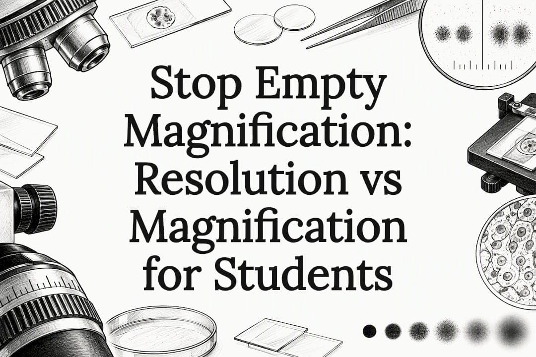
Magnification makes a specimen appear bigger; resolution determines whether you can actually see more once it does. A 20× objective paired with a 10× eyepiece gives you 200× magnification, but if the optics can't separate two nearby structures, cranking up magnification further just produces a bigger, blurrier blob. That's called empty magnification, and it happens the moment you exceed what the numerical aperture and wavelength of light can physically resolve.
TL;DR:
- Higher numerical aperture objectives, not higher magnification numbers, deliver actual improvements in image resolution and detail.
- Digital zoom enlarges pixels without enhancing optical resolution, leading to empty magnification and blurry images.
- The practical resolution limit for light microscopes is about 0.2 micrometers, determined by the objective's NA and the wavelength of light used.
- Useful magnification ranges from 500× to 1000× times the objective's NA, beyond which images only become fuzzier without added detail.
- Proper matching of objective NA, condenser alignment, and sensor pixel size is essential for achieving real microscopy detail.
Table of Contents
- Resolution vs Magnification: The Core Difference
- What Resolution Actually Measures (and How to Calculate It)
- Useful Magnification vs Empty Magnification
- Optical Resolution vs Digital Resolution: Where Sensors Come In
- Practical Examples and Quick Calculations for Your Lab Notebook
- Choosing Objectives, Cameras, and Settings for Real Detail
- Why This Distinction Changes How You Teach and Buy Microscopes
- Where Wevision Fits Into Your Next Microscope Decision
- Sources
- FAQ
Resolution vs Magnification: The Core Difference
You need both terms working correctly in your head before any formula makes sense, because students almost always mix them up. Magnification is a ratio: how much larger the image appears compared to the real object. Resolution is a performance limit: the smallest distance between two points where your microscope can still show them as two things instead of one smudge.
Total magnification = objective magnification × ocular magnification.
Here's how that plays out on your bench:
- A 20× objective with a 10× eyepiece gives you 200× total magnification.
- A 40× objective with the same 10× eyepiece jumps to 400×.
- A 100× oil immersion objective with a 10× eyepiece can provide high magnification near 1000×.
That math is simple and reliable for glass optics. Digital magnification works differently and deserves a separate mental category. When you zoom digitally on a camera feed or stretch an image on a monitor, you're enlarging pixels, not resolving new structure. The image gets bigger; the detail stays exactly where it was. This distinction matters more once you're shopping for a scope, because a spec sheet listing "2000× digital zoom" is describing screen stretching, not resolving power.
What Resolution Actually Measures (and How to Calculate It)

Resolution, sometimes called resolving power, is the minimum distance at which your microscope still shows two adjacent points as separate. It depends on two physical variables: the wavelength of light (λ) and the numerical aperture (NA) of the objective. The relationship comes from the Abbe and Rayleigh formulas, both of which boil down to the same idea: shorter wavelengths and higher NA produce finer resolution.
Numerical aperture describes how much light cone an objective can gather and focus, and it's printed right on the barrel of every objective lens, usually as a number like 0.25, 0.65, or 1.4. It's not the same as magnification. A 40× objective might have an NA of 0.65 or 0.95 depending on the manufacturer, and that NA number, not the "40×" printed next to it, determines how much detail you'll actually see.
By the numbers: With visible light at roughly 550 nanometers and a high-NA oil immersion objective near 1.4, the practical resolution limit of a light microscope settles around 0.2 micrometers. Nothing smaller than that will ever resolve as two separate points under standard light optics, no matter how much you magnify the image afterward.
Rough NA-to-resolution relationships you'll see across common objectives:
- Low-power dry objectives (NA ≈ 0.25) resolve down to roughly 1.1 μm.
- Mid-range dry objectives (NA ≈ 0.65) resolve down to roughly 0.4 μm.
- High-NA oil immersion objectives (NA ≈ 1.4) resolve down to roughly 0.2 μm.
Worth noting: computational tricks can pinpoint a single isolated point's position to a few tens of nanometers, far below these numbers. But locating one dot precisely is a different task than telling two dots apart, and it doesn't change the fundamental resolving power of the optical system.
Useful Magnification vs Empty Magnification
Every objective has a magnification range that's actually worth using, and it's tied directly to NA, not to whatever number is stamped on the eyepiece. The accepted rule of thumb, cited across microscopy education resources, puts useful magnification between 500× and 1000× the objective's numerical aperture/03%3A_Microscopy/3.01%3A_Looking_at_Microbes/3.1D%3A_Magnification_and_Resolution). Go past that ceiling and you're not gaining information anymore, you're just stretching a blur.

Empty magnification is what happens when you cross that line. The image gets larger on your screen or through the eyepiece, but no new structure appears, because the optics already hit their resolving limit. It looks like more detail is coming, right up until you realize the edges are just softer and fuzzier, not sharper.
Compare two setups:
- A ×20 objective with 0.5 NA gives a useful magnification range of roughly 250× to 500×, and its dmin (minimum resolvable distance) sits around 0.55 μm.
- A ×40 objective with 0.9 NA gives a useful range of roughly 450× to 900×, and dmin drops to around 0.3 μm.
The second setup delivers real gains, both in magnification headroom and in resolving power, because NA nearly doubled, not because the number on the barrel got bigger. This is why Nikon's own microscopy formulas resource tells lab technicians to choose objectives based on NA first and treat the eyepiece as secondary.
Pro Tip: Before buying or swapping an eyepiece, check the objective's NA number stamped on the barrel. A higher-NA objective paired with a modest eyepiece almost always outperforms a low-NA objective paired with an aggressive eyepiece, even when the final magnification number looks smaller.
Optical Resolution vs Digital Resolution: Where Sensors Come In
Once you put a camera on your microscope, a second bottleneck enters the picture: the sensor. Pixel size on the sensor, usually measured in microns per pixel, decides whether the camera can actually capture what the optics resolve. This follows the Nyquist sampling principle, which requires at least two pixels across the smallest resolvable feature to record it accurately, a standard laid out clearly in research on digital microscopy resolution.
Stretching the image on a bigger monitor afterward doesn't fix an undersized sensor. You're just displaying the same recorded pixels larger, the digital equivalent of empty magnification.
A 3.0 μm/pixel sensor without enough intermediate optical magnification to shrink that effective sample area will blur detail your objective already resolved. Vendors sometimes advertise big on-screen magnification numbers that don't match the calculated optical magnification once sensor size and tube lens differences are factored in, a gap documented in guidelines on digital microscope magnification reporting. Reporting microns per pixel instead of a marketing "×" number gives you a cleaner, vendor-neutral way to judge real image quality.
Practical Examples and Quick Calculations for Your Lab Notebook
Run these numbers before your next lab session, and you'll never be caught guessing at what your setup can actually show.
Total magnification for common objective and eyepiece pairs:
- 10× objective × 10× eyepiece = 100×
- 20× objective × 10× eyepiece = 200×
- 40× objective × 10× eyepiece = 400×
Resolution (dmin) using the Abbe relationship at λ = 550 nm:
At NA 0.5, dmin ≈ 0.55 μm. At NA 0.9, dmin ≈ 0.3 μm. At NA 1.4 (oil immersion), dmin ≈ 0.2 μm, matching the practical optical resolution limit cited earlier.
For a 0.9 NA objective, the useful magnification range runs from about 450× to 900×, following the 500 to 1000 times NA guideline. Anything you push beyond 900× on that objective is empty magnification.
Three-step checklist for your notebook:
- Record the NA printed on each objective you use, not just its magnification number.
- Note your camera's sensor pixel size in microns before recording images.
- Divide dmin by pixel size to confirm you meet the two-pixel Nyquist minimum.
Choosing Objectives, Cameras, and Settings for Real Detail
Numerical aperture should drive every lens decision you make, with magnification treated as a secondary convenience. Immersion oil raises NA meaningfully on compatible objectives, which is why 1.4 NA oil objectives outresolve dry lenses at the same or even higher stated magnification. On the camera side, match sensor pixel size to your optical dmin using the Nyquist guidance above, and resist the urge to lean on digital zoom once you've hit your optical ceiling.
Quick setup checklist:
- Pick objectives by NA rating first, magnification number second.
- Use immersion oil on compatible high-NA objectives for fine specimens.
- Confirm condenser alignment; a misaligned condenser wastes NA you already paid for.
- Set camera pixel size and binning to satisfy Nyquist sampling for your target dmin.
Pro Tip: A well-aligned condenser matched to your objective's NA can improve resolved detail more than swapping to a pricier eyepiece. Check condenser NA against objective NA before assuming you need new optics.
Why This Distinction Changes How You Teach and Buy Microscopes
Students who grasp resolution vs magnification stop chasing bigger numbers and start asking better questions about NA and sample prep. Wevision builds its setup guides and video demonstrations around that exact shift, because a well-matched instrument teaches the physics on its own, one clear image at a time.
— Oliver
Where Wevision Fits Into Your Next Microscope Decision
Wevision builds premium compound microscopes with glass optics chosen for real resolving power, not inflated magnification claims on a box. Every model ships with instructional video demonstrations and setup guides that walk you through condenser alignment, objective selection, and illumination, the exact variables that determine whether your students see structure or just a bigger blur.
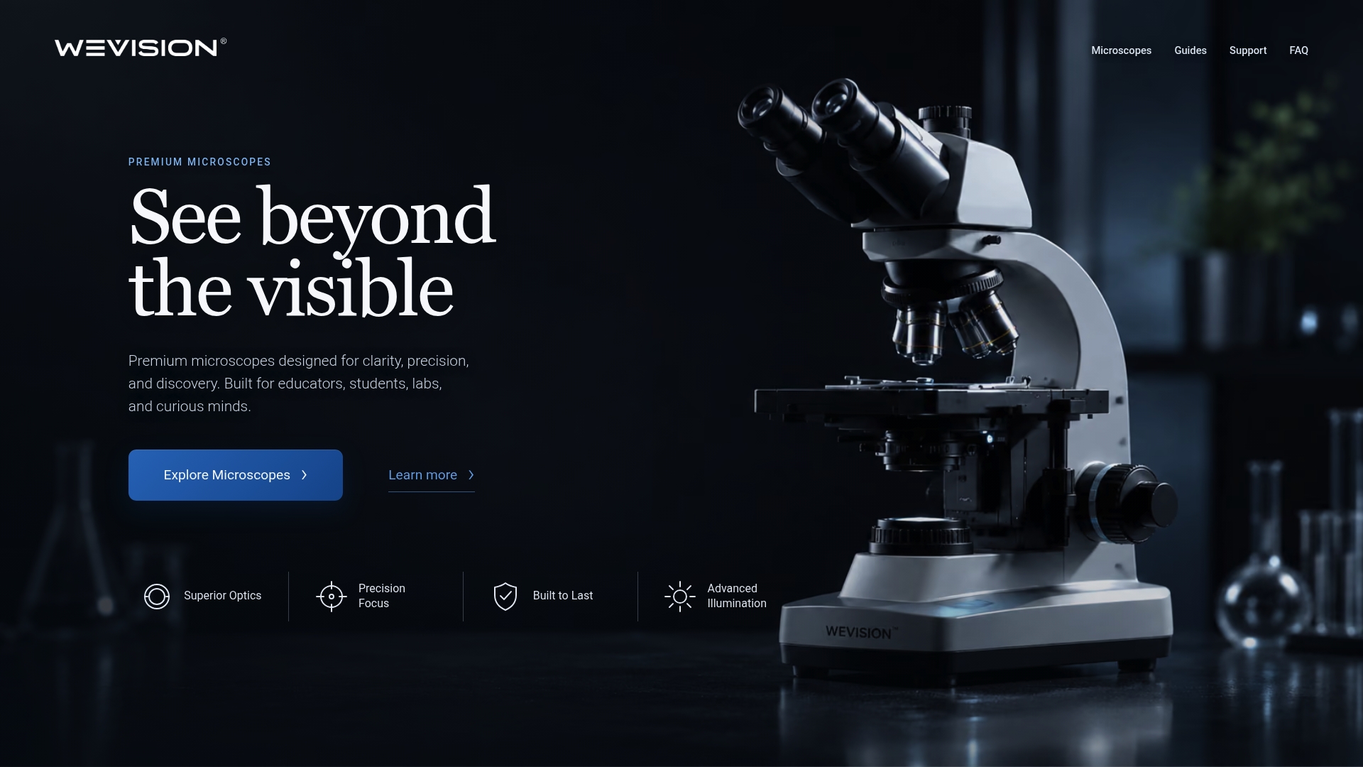
If you're outfitting a classroom or lab bench, the Wevision microscope lineup covers a range of objective and NA combinations suited to coursework from basic cell biology through more demanding specimen work. The Wevision M2 Monocular Compound Microscope, covering 40X to 2000X, and the Wevision M1 Binocular Compound Microscope, covering 40X to a high magnification level, both pair usable magnification ranges with the optics needed to make that range meaningful and not empty. Responsive email support backs every purchase, so setup questions about NA, immersion oil, or camera pairing don't sit unanswered. Browse the full Wevision catalog and pick the model that matches the resolution your coursework actually demands.
Sources
- Microscopy chapter (NCBI Bookshelf)
- Relationship between magnification and resolution in digital microscopy (PMC)
- Useful magnification range (Nikon / FSU resource)
FAQ
Does magnification affect resolution?
No. Magnification changes how large an image appears, while resolution depends on the objective's numerical aperture and the wavelength of light used. You can magnify an image well past your microscope's resolution limit, but you'll only be enlarging blur, a state known as empty magnification.
What does 20x magnification mean on a microscope?
A "20×" label on an objective means that lens alone makes the specimen appear 20 times larger than its actual size. Paired with a standard 10× eyepiece, total magnification reaches 200×, calculated by multiplying objective magnification by ocular magnification.
Is lower resolution better for microscopes?
No, lower resolution means the microscope can distinguish fewer fine details, since resolution measures the smallest separable distance between two points. Higher resolving power, tied to higher numerical aperture and shorter wavelengths, is what lets you see finer structure, down to about 0.2 micrometers with top oil immersion objectives.
Is 10x or 40x bigger?
A 40× objective produces four times more magnification than a 10× objective when paired with the same eyepiece, so it enlarges the specimen more. But bigger magnification alone doesn't guarantee sharper detail. That depends on the numerical aperture of each specific 40× objective, which is why Wevision's compound microscope lineup lists NA alongside magnification for every model.
Why does my microscope image look blurry at high magnification?
Blur at high magnification almost always means you've exceeded your objective's useful magnification range, generally 500× to 1000× times its numerical aperture. Switching to a higher-NA objective, rather than adding more eyepiece or digital zoom, is the fix.