

You need a brightfield compound microscope with a 100x oil immersion objective, giving you 1000x total magnification, to read a Gram stain correctly. The stain itself sorts bacteria into two camps within minutes: Gram-positive organisms hold onto crystal violet and look purple, while Gram-negative organisms lose it and pick up safranin, turning pink. Add in cell shape and clustering pattern, and you get a same-day presumptive read long before a culture ever finishes growing.
TL;DR:
- Accurate Gram stain reading requires a 100x oil immersion objective for clear resolution of bacteria.
- Proper timing of decolorization, using controlled rinse rather than exact seconds, is critical to prevent false Gram-positive or Gram-negative results.
- Thin, well-prepared smears with fresh reagents and well-maintained microscopes significantly improve identification accuracy.
- Neutrophil visibility and shape are vital quality checks that indicate whether the stain technique is reliable.
- Budget microscopes often lack the optical quality needed for dependable results; high-quality optics and correct illumination systems are essential for trustworthiness.
Table of Contents
- What Microscope and Accessories You Need for Gram-Stain Viewing
- The Four-Step Gram Stain Procedure, With Timings That Matter
- How to Focus and View a Gram-Stained Slide Under Oil Immersion
- Reading the Slide: Color, Shape, and What They Tell You
- Fixing the Most Common Gram Stain Mistakes
- Safety and Preparation Basics You Shouldn't Skip
- Getting Your Setup Ready for Oil Immersion Work
- A Few Practical Habits Worth Building Early
- Why Your Microscope Choice Shapes Every Gram Stain Result
- Where to Go for Deeper SOP Detail
- Sources
- FAQ
What Microscope and Accessories You Need for Gram-Stain Viewing
You cannot read a Gram stain with a standard classroom scope maxed out at 400x. Bacteria are simply too small to resolve without immersion oil at 1000x total magnification, which means your setup needs a 100x oil immersion objective paired with 10x oculars. Skip the oil and you get a hazy, low-contrast smear instead of crisp purple rods or pink cocci.
Beyond the objective itself, a few features separate a frustrating session from a smooth one:
- A compound brightfield microscope with a 100x oil immersion objective and 10x oculars (1000x total).
- An adjustable condenser and iris diaphragm so you can properly align illumination (Koehler alignment gives you even, glare-free light across the field).
- A mechanical stage with fine focus control, since oil immersion work leaves almost no room for hand-jitter.
- Immersion oil formulated for microscopy (never substitute a random household oil).
- Lens cleaner and lens paper to remove oil residue after every session.
- Bibulous paper for blotting slides without smearing the smear.
Before you ever load a slide, run a quick check: clean optics, fresh oil bottle with no bubbles, condenser centered, and stage clear of old oil smudges. That thirty-second habit prevents most of the focusing headaches beginners blame on "bad slides."
The Four-Step Gram Stain Procedure, With Timings That Matter
Gram staining is a four-reagent sequence, but the timings are where technique either holds up or falls apart. NCBI's StatPearls entry on Gram staining stresses that sample quality and a thin, even smear matter as much as the reagents themselves, so start there.
- Prepare and fix the smear. Spread a thin film of your specimen on a clean slide and let it air dry completely. Fix it either by passing it briefly through a flame (heat fixation) or by flooding it with methanol for about a minute. Methanol tends to preserve cell morphology better and cuts down on aerosol risk compared to heat, which is why many current lab procedures favor it when it's available.
- Apply crystal violet (primary stain). Flood the smear for roughly 60 seconds, then rinse gently with water.
- Apply Gram's iodine (mordant). This locks the crystal violet into Gram-positive cell walls. Leave it on for about 60 seconds, then rinse.
- Decolorize. This is the step that makes or breaks your slide. Apply the decolorizer (acetone-alcohol or alcohol alone) for as little as 3 to 15 seconds, or simply until the runoff turns clear rather than purple. Watching the runoff color matters more than watching the clock.
- Counterstain with safranin (or basic fuchsin). Apply for 30 to 60 seconds, rinse, then blot gently with bibulous paper. Never rub the slide.
Let the slide air dry fully before you touch it with oil. Rushing the drying step is one of the most common reasons a technically correct stain still looks smeared under the objective.
How to Focus and View a Gram-Stained Slide Under Oil Immersion
Start on low power, not high. Scanning at low magnification first lets you locate a representative, evenly stained region before you commit to oil, and it saves you from smearing oil across a field that turns out to be too thick to read anyway.
Here's the sequence that actually works:
- Scan at 10x or 40x to find a thin, single-cell-layer area, ideally one containing neutrophils if you're reading a clinical specimen.
- Center that field, then switch to the 40x objective to confirm focus and framing.
- Rotate the nosepiece away from the slide, place one drop of immersion oil directly on the target spot, and swing the 100x objective into the oil without shifting the stage.
- Fine-focus only. Moving the stage after the oil is applied smears the oil and risks lens contamination, so resist the urge to hunt around once you're in oil.
- Adjust the condenser and iris diaphragm for even illumination rather than cranking up brightness, which just washes out contrast.
Pro Tip: If you're photographing the field for a report or a teaching file, take the image after you've confirmed the read with your own eye. A camera sensor can misrepresent subtle purple-versus-pink shading, and your eye at the eyepiece is still the more reliable judge.
Reading the Slide: Color, Shape, and What They Tell You
Color tells you cell-wall chemistry. Gram-positive organisms retain the crystal violet and iodine complex, appearing deep purple, while Gram-negative organisms lose that complex during decolorization and take up the pink safranin counterstain instead. Shape and arrangement tell you the rest of the story.
- Cocci in clusters often suggest staphylococci; cocci in chains point toward streptococci.
- Rod-shaped organisms (bacilli) can be Gram-positive or Gram-negative, and their size and grouping narrow the possibilities further.
- Fields with intact neutrophils double as a built-in quality check. If the neutrophils themselves stain a clean purple or pink and organisms inside them are visible, your technique held up.
Spend at least a minute scanning several representative fields rather than judging the whole slide from one lucky patch. A single field can mislead you, especially on a specimen from a nonsterile site where mixed flora is expected rather than abnormal. On specimens from normally sterile sites, like blood or spinal fluid, seeing any organisms at all is itself a significant finding worth flagging for further testing.
Not everything shows up reliably. Spirochetes, mycobacteria, and other unusually small or thin-walled organisms often stain poorly or not at all, so a clean Gram stain never rules them out. When clinical suspicion points that way, an acid-fast stain or dark-field microscopy is the next step, not a repeat Gram stain.
Fixing the Most Common Gram Stain Mistakes
Most bad Gram stains trace back to one of a handful of repeatable errors, and the decolorization step causes more misreads than any other part of the procedure.
- Over-decolorization strips crystal violet even from Gram-positive cells, making them falsely appear pink. Watch the runoff color instead of counting seconds blindly, and stop the moment it clears.
- Under-decolorization leaves Gram-negative cells looking falsely purple. If every organism on the slide reads Gram-positive with no exceptions, suspect this before you suspect the specimen.
- Smears that are too thick trap stain and distort colors regardless of timing; thin the smear next time and pick a clearer area to read this time.
- Rough blotting or old cultures can strip cells off the slide entirely or distort their shape, so blot gently and prefer fresh growth when you have a choice.
- Run a known control slide (a mixed Gram-positive and Gram-negative culture) periodically to confirm your reagents and technique are both still reliable before you trust a diagnostic read.
Safety and Preparation Basics You Shouldn't Skip
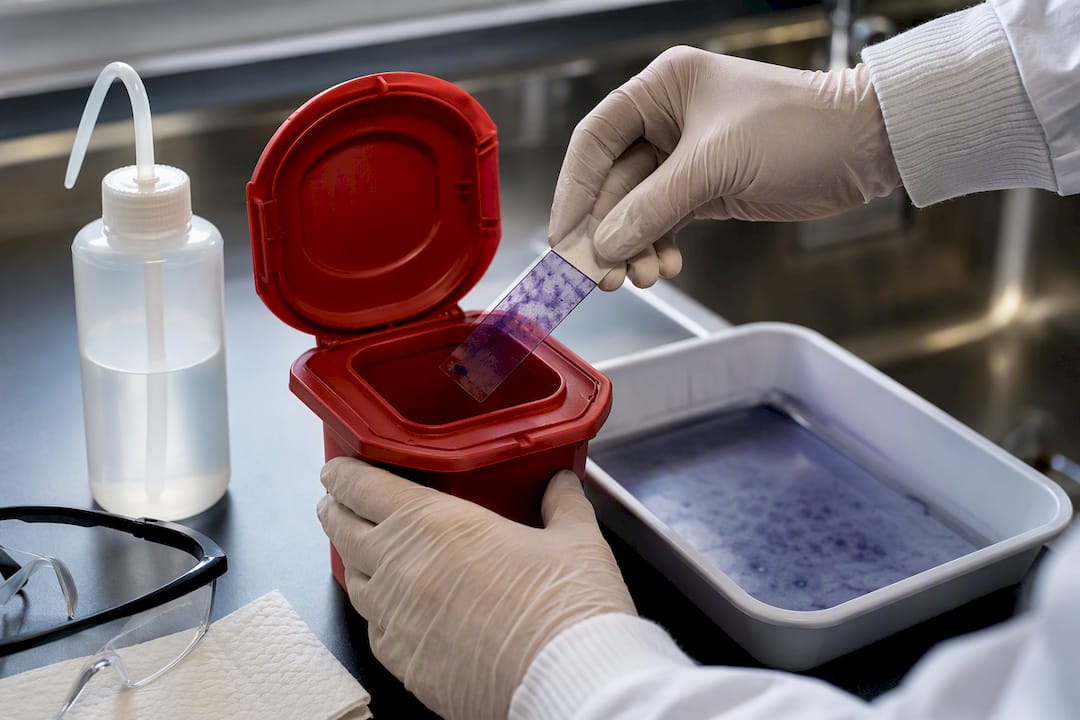
Wear gloves and eye protection when handling reagents, and work over a sink or tray in case of spills. Use a biosafety cabinet for specimens with unknown or higher-risk pathogens. Choose methanol fixation over heat when morphology preservation and lower aerosol risk both matter, dispose of stained slides and waste per your lab's protocol, and clean oil off your objective immediately after use to keep it from drying into a hard film.
Getting Your Setup Ready for Oil Immersion Work
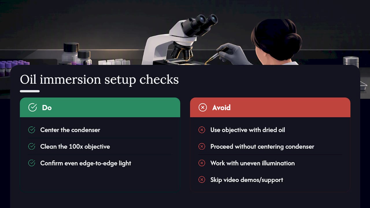
Before your first slide, confirm your condenser is centered, your 100x objective is clean and free of dried oil, and your light source gives even illumination edge to edge. Wevision's compound microscopes come with video demonstrations that walk through this exact alignment process, along with responsive support if something looks off. [Author credential placeholder: insert Oliver's relevant microscopy or laboratory training background here]
A Few Practical Habits Worth Building Early
Scan for neutrophils before you judge anything else. They tell you almost instantly whether your technique held up, since a poorly decolorized field usually shows itself in how those cells stain, too. Watch the decolorizer runoff rather than trusting a mental stopwatch. Beyond that, this comes down to repetition: read a few dozen slides deliberately, compare your calls against a known control, and the interpretation skills stop feeling like guesswork within a few sessions.
— Oliver
Why Your Microscope Choice Shapes Every Gram Stain Result
The reagents matter, but the optics decide whether you can actually trust what you're looking at. A dim condenser, a scratched objective, or oil that spreads because the stage got bumped will wreck a technically perfect stain before you even finish reading it. Wevision builds premium compound microscopes around exactly this problem: glass optics, oil-immersion compatible objectives, and illumination systems designed to hold up at 1000x without the guesswork that comes with budget scopes. These models often include video demonstrations covering setup and Koehler alignment, plus email support if your slide still looks off after you've followed the steps here. If you're outfitting a classroom, a teaching lab, or your own bench, visit the Premium microscopes page to see the current lineup and get the setup resources you'll actually use.
Where to Go for Deeper SOP Detail
- Rice University's Gram stain protocol and technique notes
- NCBI StatPearls: Gram Staining
- CDC Routine Microscopy Student Laboratory Exercises (Gram stain job aid)
- Cleveland Clinic: Gram Stain overview
This article is general information, not a substitute for advice from a qualified doctor. Consult a qualified healthcare professional about your own circumstances before acting on anything here.
Sources
- Rice University — Gram stain (BIOL/BIO labs resource)
- Cleveland Clinic — Gram Stain: What It Is, Purpose, Procedure & Results
- Gram Staining - StatPearls - NCBI Bookshelf - NIH
- CDC — Routine Microscopy Student Laboratory Exercises (Gram stain job aid)
FAQ
What Can a Gram Stain Tell You?
A Gram stain shows whether bacteria are Gram-positive (purple) or Gram-negative (pink), along with their shape and how they cluster together. That combination gives a presumptive identification hours before a culture would confirm it, which often guides an initial choice of antibiotic therapy.
What Are the Four Basic Steps of Gram Staining?
The four steps are crystal violet (primary stain), Gram's iodine (mordant), a decolorizing agent, and safranin or basic fuchsin (counterstain), with a water rinse between each. Decolorization is the step to watch most closely, since it's the most common source of a misread slide.
What Does It Mean If a Gram Stain Is Abnormal?
An abnormal result usually means bacteria appear where none are expected, such as in a normally sterile specimen like blood or spinal fluid. It can also mean a mix of organisms shows up on a specimen where a single dominant type was expected, which sometimes points to contamination or a technical decolorization error rather than a true polymicrobial infection.